Congenital Muscular Dystrophy (CMD)
Types Of Congenital MD
At least 30 different types of CMD are now recognized (see the Types of CMD chart). At first glance, the various types of CMD seem to have little in common other than their early onset. But on the molecular level, the types can be grouped how their faulty protein affects cells.
A very small group of CMDs are linked to proteins that affect what happens inside muscle fibers, affecting how the fibers process signals from the nervous system, for example, or how they handle calcium.
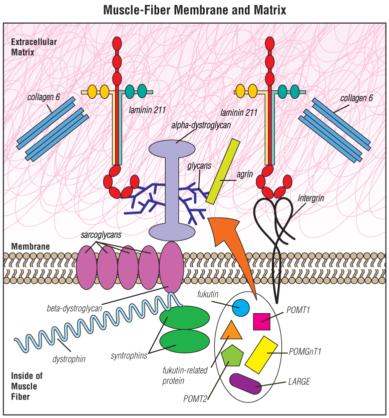
But the vast majority of CMD types are related to proteins that make up or interact with the extracellular matrix that surrounds muscle fibers (see CMD: The Cellular View below).
Several types of CMD that arise from gene mutations that initially seemed unrelated now appear to be related to defects in proteins that "sugar-coat" (glycosylate) a matrix protein, allowing it to connect with other proteins.
The extracellular matrix and CMD
The extracellular — outside the cell — matrix is the substance that surrounds the cells of a tissue, such as muscle, providing physical and biochemical support.
An important role of the matrix around muscle fibers is force transmission. For a muscle to pull against bones, it needs to have contact with something that transmits force from the muscle fibers onto the tendons and bones.
When all is going well, the matrix transmits that force, as well as chemical signals that muscles need to stay healthy.
The matrix is a key supporting structure for the survival and regeneration of muscle. When cells lose touch with their surrounding matrix — as happens in most types of CMD — trouble follows.
CMD: The cellular view
Looking for more information, support or ways to get involved?
Find MDA
in your Community
-

Grants at a Glance
Read More -

Search for Clinical Trials
Learn More

